
Which approach reveals more about your client's health concerns?
Story at a Glance:
• Complementary Approaches: Visual inspection and palpation each reveal different aspects of a client's health—together they create a comprehensive diagnostic picture.
• Visual Clues: Changes in color, texture, and morphology of the ear can indicate past, present, or future health conditions even before symptoms appear.
• Tactile Confirmation: Tender points often reveal issues that aren't visible, helping practitioners pinpoint the most effective treatment areas.
• Individual Variation: In stressed or depleted clients, one method may yield more information than the other, making proficiency in both essential.
• Enhanced Results: Combined diagnostic approaches lead to more precisely targeted treatments and improved client outcomes.
Introduction: Two Paths to Creating Effective Auricular Protocols
As auriculotherapy practitioners, we face a fundamental question with each new client: should we trust what we see or what we feel? Should we rely on visual inspection of the ear or tactile feedback through palpation to guide our treatment protocols?
The answer, as experienced practitioners know, isn't either/or—it's both.
A Recent Case Study: The Invisible Heel Pain
We were recently at the Hemp for Humanity Summit in Sedona where we were delivering ear seeding sessions to summit attendees. One attendee complained of right-sided heel pain, but during our initial examination, there were no visible auricular signs in the heel area of her ear.
View this post on Instagram
However, when Elie performed palpation, he found significant tenderness on the heel point on her right ear. This case perfectly illustrates an important principle: when auricular signs are visibly present, there's a very high chance of past, present, or future issues in that corresponding body area.
But when no visual signs appear, palpation becomes the crucial next step in diagnosis.
The Visual Journey: Auricular Diagnosis Through Inspection
Imagine the ear as a living map, continuously updating to reflect what's happening throughout the body. Visual inspection—the systematic observation of the ear's appearance—can reveal functional disorders and mental-emotional conditions that conventional diagnostic methods might miss entirely.
When examining the ear, look for these visual indicators:
Skin Alterations
#1 Color Changes:
Areas of redness (hyperemia) often signal acute conditions or inflammation, while paleness might indicate deficiency or chronic issues. A white discoloration at the thyroid point could suggest hypothyroidism, while brown dots on mammary points might point to blood stagnation in the breast.
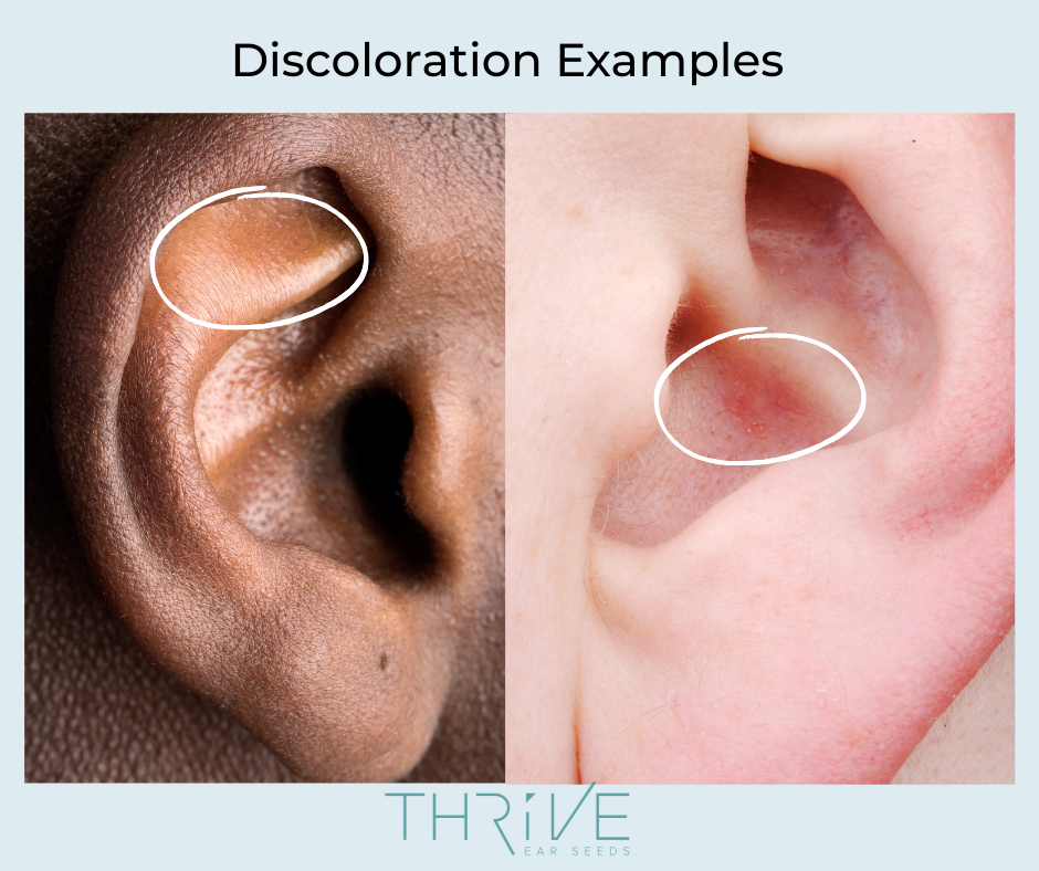
#2 Vascular Networks:
Small, dilated blood vessels (telangiectasia) can indicate underlying issues in the corresponding body area.
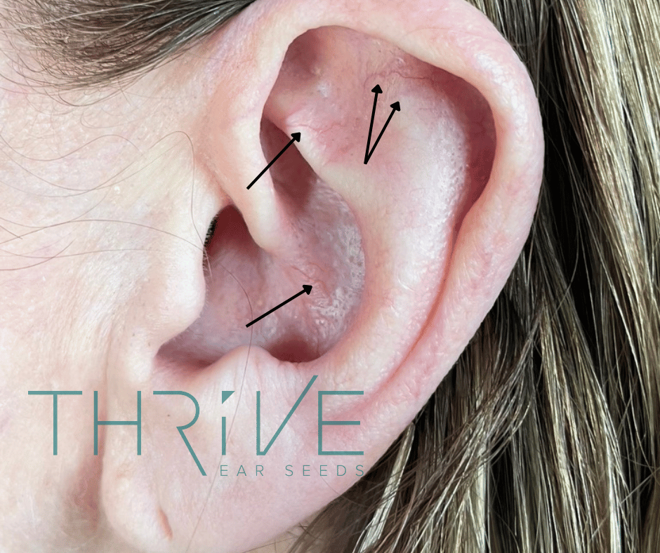
#3 Texture Changes:
Pay attention to flakiness, roughness, bumpiness, or scaliness. Indentations or depressions might suggest swelling or edema in the corresponding body part.
#4 Growths and Protrusions:
Observe any nodules or papule-like formations. A hard white protrusion at the stomach area may be associated with digestive issues like acid reflux, while a drooping area of the helix at the apex may indicate allergies.

#5 Lines and Grooves:
Creases like the brain line may indicate cognitive issues or the myopia groove might indicate vision problems, while the tinnitus line may be associated with ear ringing or hearing loss.
#6 Press Marks:
During palpation, notice if press marks remain after removing the probe—their absence might suggest the point is free of swelling.
A Tale of Two Back Pain Cases
During our sessions at the Hemp for Humanity Summit in Sedona, Robin treated two attendees with low back pain who perfectly illustrated the complementary nature of visual and tactile diagnosis.
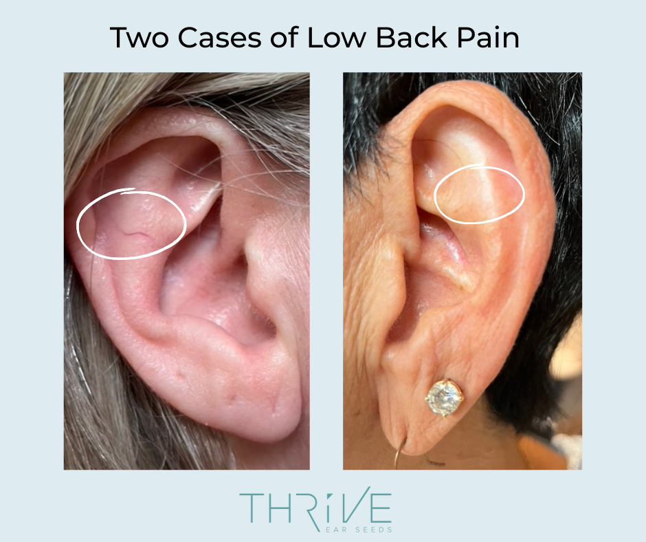
The first client, as shown on the left in the photo above, had a clearly enlarged blood vessel in the low back region of her ear—a visible sign that aligned with her reported history of chronic back issues.
When Robin performed palpation, she found tender spots directly on the blood vessel and in other ear regions corresponding to low back pain, confirming the visual diagnosis.
The second client, shown on the right, presented with no visible auricular signs despite similar complaints.
Yet upon palpation, she exhibited significant tenderness in the low back region of her ear, and the probe left noticeable indentations—indicating swelling or edema in the corresponding low back area that wasn't detectable through visual inspection alone.
These two cases, treated side by side, demonstrated how both diagnostic approaches can unveil different aspects of the same condition.
Morphological Features
The ear's overall shape and structure can provide valuable context. Anthropologists have documented tremendous variation in ear morphology, with some characteristics having hereditary tendencies.
For example, the proportion of the earlobe to the entire ear is sometimes observed in relation to longevity—a larger earlobe potentially indicating a longer life. Cartilage deformities may be particularly relevant when examining disorders of the spine and skeletal system.
Spotlight: Frank's Sign and Heart Health
One of the most fascinating examples of auricular diagnosis in Western medicine is Frank's sign—a diagonal crease across the earlobe that starts near the intertragic notch.
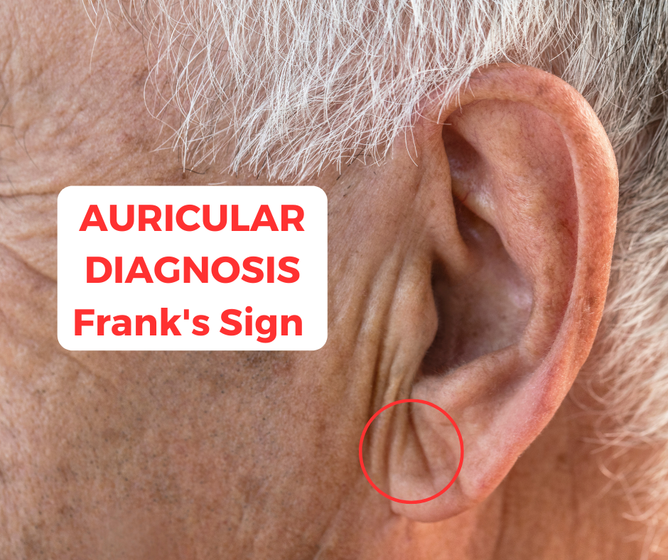
Since Dr. Sanders T. Frank described it in 1973 as an "aural sign of coronary artery disease," numerous studies have explored its correlation with cardiovascular risk. A shallow groove might signify arrhythmia, while a deeper groove could indicate higher risk of coronary artery disease.
Frank's Sign: Beyond the Obvious
One of Robin's clients presented with a clear heart line (Frank's sign) across his earlobe. When questioned about his cardiovascular health, he noted that while recent testing had not revealed any signs of coronary artery disease, he did have a family history of mitral valve prolapse and was being regularly monitored by his doctor.

This case illustrates how auricular signs can sometimes reflect genetic predispositions or future risk factors rather than current conditions—highlighting the value of these visual indicators for preventative health awareness and early monitoring.
The Tactile Journey: Palpation as "Listening" Through Touch
While visual inspection gives us the overview, palpation helps us pinpoint active treatment points with precision. Using pressure to identify tender areas can reveal corresponding imbalances in the body that might not be visually apparent.
The Art of Palpation
Effective palpation requires skill, sensitivity, and the right tools—typically a metal probe with a round, blunt tip approximately 1.5mm in diameter.
A Step-by-Step Approach to Ear Palpation
Let's walk through the process using low back pain as an example:
1. Hold the probe like a pen in your dominant hand.
2. With your non-dominant hand, gently stretch and support the auricle to access all surfaces.
3. Use gentle but firm pressure perpendicular to the ear's surface. Glide smoothly and maintain even pressure for accurate comparison. When testing a particular area, like the low back, it can be helpful to lift the probe and press between spots.
4. Watch for Reactions: Pay close attention to the client's non-verbal cues—a grimace, blink, or twitch (the 'grimace sign') can be more reliable than their verbal feedback (watch the heel pain video above to see a client reaction!).
5. Check for Alignment: When you find a tender point, check if there are other tender points aligned with it, following Nogier's alignment principle.
Pro Tip: When Ears Don't Respond
Some clients, despite having obvious pain or health issues, will not have any tenderness when you're palpating the ear. This may be due to extremely high stress levels potentially causing the nervous system to be in a sympathetic dominant state.
In other clients that are physically and/or energetically depleted, they may not have enough energy or qi to notice tenderness upon palpation. In these cases, you'll rely more on visual inspection and point locations to develop your protocol.
This reinforces why mastering both diagnostic approaches is essential—when one method yields limited information, the other becomes your primary guide.
Hidden Signs Revealed Through Touch
In a fascinating demonstration of how palpation can reveal what the eye might miss, Elie once examined his employee, Jess, who had been diagnosed with hypothyroid. He wanted to see if she had any auricular signs that indicated this condition.
Initially, visual inspection didn't reveal much in the way of auricular signs. However, as he palpated the thyroid area of her ear with the back of his tweezers, he felt the instrument slide over a distinct bump! It was a protrusion that had been difficult to see but through palpation was plain as day.

Jess confirmed that the area was tender, providing further confirmation of an imbalance in the thyroid. This case perfectly illustrates how tactile examination can detect subtle morphological changes that visual inspection alone might miss—especially in areas where the ear's natural contours can obscure small protrusions.
The Synergy Approach: Combining Visual and Tactile Diagnosis
The true magic happens when we integrate both diagnostic approaches. Visual cues can direct our palpation efforts to promising areas, while tactile feedback confirms and refines our visual observations.
This comprehensive methodology allows you to:
- Validate findings through multiple channels
- Identify points that might be missed by using only one approach
- Move beyond standardized protocols to create truly personalized treatments
Rapid Relief: When Both Approaches Lead to Breakthrough
Remember the second client with low back pain mentioned earlier—the one who had no visible auricular signs but showed tenderness and indentation upon palpation? Her story continued with remarkable results.
She approached Robin the very next day at the summit, excitedly reporting that her back felt significantly better after just one ear seeding session. She hadn't needed to use her heating pad that night—a regular necessity before the treatment—and when she woke up the following morning, the pain was almost completely gone.
This dramatic improvement underscores the power of combining diagnostic approaches. Without palpation, the reactive points might have been missed entirely, as they showed no visible signs. Yet by carefully detecting tenderness and tissue changes through touch, Robin was able to precisely target the treatment, leading to rapid, effective relief.
Conclusion: The Art of Integration
Rather than debating which method is superior, the most effective practitioners develop fluency in both visual and tactile diagnosis. By combining these complementary approaches with a strong knowledge of point locations, you can create truly tailored treatments that address each client's unique presentation.
This integrated approach isn't just more thorough—it's the key to unlocking auriculotherapy's full therapeutic potential.
Your Journey to Mastery: A Personal Note
Practice is essential for mastering both diagnostic skills. For auricular diagnosis, it's helpful to see pictures of the auricular signs because they present slightly differently on each person's ear.
Our ear maps are a guideline—they're fluid and shift a little on each person's ear. That's why we recommend taking one of our auricular diagnosis classes to gain experience and highlight what you can look for.
You can also get access to all of our auricular diagnosis classes when you join Ear Seeds Pro Club, the ultimate resource for auriculotherapy. For true mastery and confidence building, we recommend our Ear Seeds Mastery Certification.
With practice, these skills can add immense value to your sessions and help you create customized auricular protocols to address hundreds of conditions. The journey to becoming proficient in both visual and tactile diagnosis takes time—but the results you'll achieve with your clients make every moment of learning worthwhile.
About the Authors

Robin Ray Green is a pediatric health expert, author, speaker, and business coach who co-founded the Thrive Ear Seeds Educational Platform. A prominent figure in pediatric acupuncture, Robin has shared stages with healthcare luminaries including Dr. David Perlmutter, Jack Canfield, and Dr. Elisa Song. She authored the Hay House best-seller "Heal Your Child From The Inside Out: The 5-Element Way to Nurturing Healthy, Happy Kids," offering practical solutions for children's health issues. She has been practicing auriculotherapy for over 20 years and teaches our pediatric auriculotherapy and techniques classes.
Elie Goldschmidt is an internationally recognized auriculotherapy expert who founded EarSeeds.com, Ear Seeds Academy, and co-founded Thrive Ear Seeds. Trained by Dr. Lichun Huang ("Mother of Auriculotherapy") and Dr. Raphael Nogier (son of auriculotherapy pioneer Dr. Paul Nogier), Elie has taught thousands of students. His gift lies in making complex concepts accessible and enjoyable to learn. Since launching his innovative Ear Seeds Certification in 2016, he has revitalized this ancient practice, making it more practical for real-world application.
Together, they're dedicated to making auriculotherapy accessible to both practitioners and families seeking natural health solutions.
Comments